Emiliania huxleyi Coccoliths
Jeremy Young
Palaeontology Dept.
The Natural History Museum
London, SW7 5BD
Great Britain.
Email: j.young@nhm.ac.uk
This page describes Ehux coccoliths, i.e. the characteristic
calcareous plates the cell produces. People often loosely refer to
Ehux as a coccolith, which is strictly speaking incorrect. This is
not drastically important but the correct terminology is:
Coccolithophore - the organism, including the cell and external
coccoliths.
Coccosphere - the more or less spherical layer of coccoliths
surrounding the cell.
Coccolith - the individual calcareous plates.
Ehux produces coccoliths that are approx. 2.5 x 10^-6 metres in
diameter, and which weigh approx. 1.8 x 10^-12 g each. Of this weight, the
average contents of carbon, oxygen and calcium atoms in the coccolith CaCO3
have been measured to be 0.28, 0.87 and 0.67 x 10^-12 g per coccolith
respectively (Fagerbakke et al, 1994).
For comparison, the wavelength of visible light is in the range 0.4-0.7 x
10^-6 metres, and a pinhead is about 2000 x 10^-6 metres in diameter.
[click on the small pictures on this page to view them at full size]
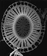
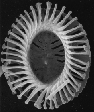
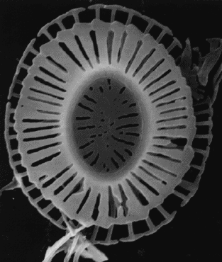
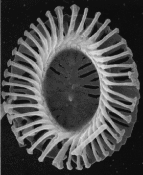
Three SEMs of E. huxleyi coccoliths: the first two of the under-side of a
coccolith (the side lying closest to the cell surface), the third from
'above' (pictures from Jeremy Young).
The coccoliths are produced inside the cell (see also
cell page)
then extruded to the outside. The coccoliths themselves are not living
structures but dead mineral structures, analogous to human bone or
fingernails, or the shells of oysters and other molluscs. Such biominerals
are intriguing structures and study of their formation, or
biomineralization, is a challenging interdisciplinary science which seeks
both to understand natural structures and processes and to learn from nature
how complex mineral structures can be formed. Coccoliths are one of the
more remarkable types of biominerals owing to the very precise control by
the organic system of every aspect of inorganic mineral growth. For this
reason and because coccolithophores are easily studied in the laboratory
coccolith formation, particularly in Ehux has been intensively
studied.
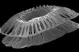
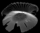
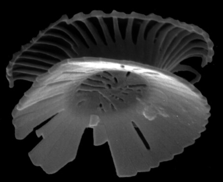
Two side-views of coccoliths. Star Trek? (Pictures from Jeremy
Young).
Coccolith shape and arrangement on the cell: Ehux coccoliths
have a very characteristic shape (placolith morphology for terminological
tyros) which has been variously likened to rivets, collar studs, and
cable-reels. There is a central tube from which two flanges or shields
extend. The shields are curved to match the curvature of the cell wall so
there is a clear top and bottom to the coccolith. The concave side of the
coccolith is nearer to the cell and is termed the proximal or lower surface,
as opposed to the upper or distal surface. The area inside the tube is open
on the distal side but filled on the proximal side by a grill. On the
coccosphere the shields of the individual coccoliths overlap and interlock
forming a robust structure.
This seems to be a self-organising effect - we believe individual coccoliths
are pushed out by the cell the right way-up, but otherwise more or less at
random into the coccosphere. Extra-cellular polysaccharide almost certainly
helps keep the coccoliths attached to the cell and random shuffling between
them results in the interlocking. This irregular self-organising structure
to the coccosphere means that the number of coccoliths per cell does not
need to be regulated closely. Typically there is a single layer of about 10
coccoliths around the cell, but some cells accumulate multi-layered
coccospheres with hundreds of coccoliths. Conversely in cultures of
Ehux it is quite common to see cells with incomplete coccospheres, or
with no coccoliths at all.
Coccolith structure and growth: coccolith formation occurs inside the
cell and has been studied both by looking at sections through cells with
coccoliths at various stages of growth and by looking at isolated
incompletely formed coccoliths. Growth starts with nucleation of an
elliptical ring of simple calcite crystals - a proto-coccolith ring. This
proto-coccolith ring is situated at what will become the base of the tube.
Growth then progresses simultaneously in various well-constrained
directions: outwards to form the proximal shield; inwards to form the
central grill; upwards and then outwards to form an outer layer of the tube
and the distal shield; obliquely upwards to form the inner layer of the
tube. This growth occurs entirely by enlargement of the crystals of the
proto-coccolith ring, with no formation of new crystals. Also each of the
developing crystal-units produces elements in each of the growth directions,
thus developing a very complex final form - particularly when contrasted to
the rhombohedral and prismatic crystals produced during inorganic calcite
growth.
One result of the complex shape is that the crystals interlock with each
other, particularly due to the difference in obliquity of the inner and out
layers of the tube. This interlocking gives the structure considerable
strength - Ehux coccoliths normally stay intact even during quite
robust laboratory preparation techniques. In addition the complex
crystal-unit structure provides the best morphological criteria for
determining the evolutionary relationships of different coccoliths.
Coccoliths formed by species closely related to Emiliania huxleyi
have the same basic structure of four elements formed from a single
crystal-unit. Ehux is characterised by the delicate hammer-like form
of the distal shield elements, with openings between each element. By
contrast most related species have solid distal shields and may have other
distinguishing features, for example in Gephyrocapsa species there is
bridge across the central area.
Coccolithogenesis: Alison Taylor has produced a
wonderful
time-lapse video
showing another coccolithophore (Coccolithus pelagicus) building its
coccoliths inside the cell and then extruding them out onto the cell
surface. The coccoliths are rather large relative to the cell size; if
scaled up to human size it would be like a person giving birth to a car
wheel or a dustbin lid.
For more detailed and specialised information on the geometry and formation
of the coccoliths, click
here.
References
Ehux
home page